

How TMS Works: The Neuroscience of Magnetic Stimulation for Depression

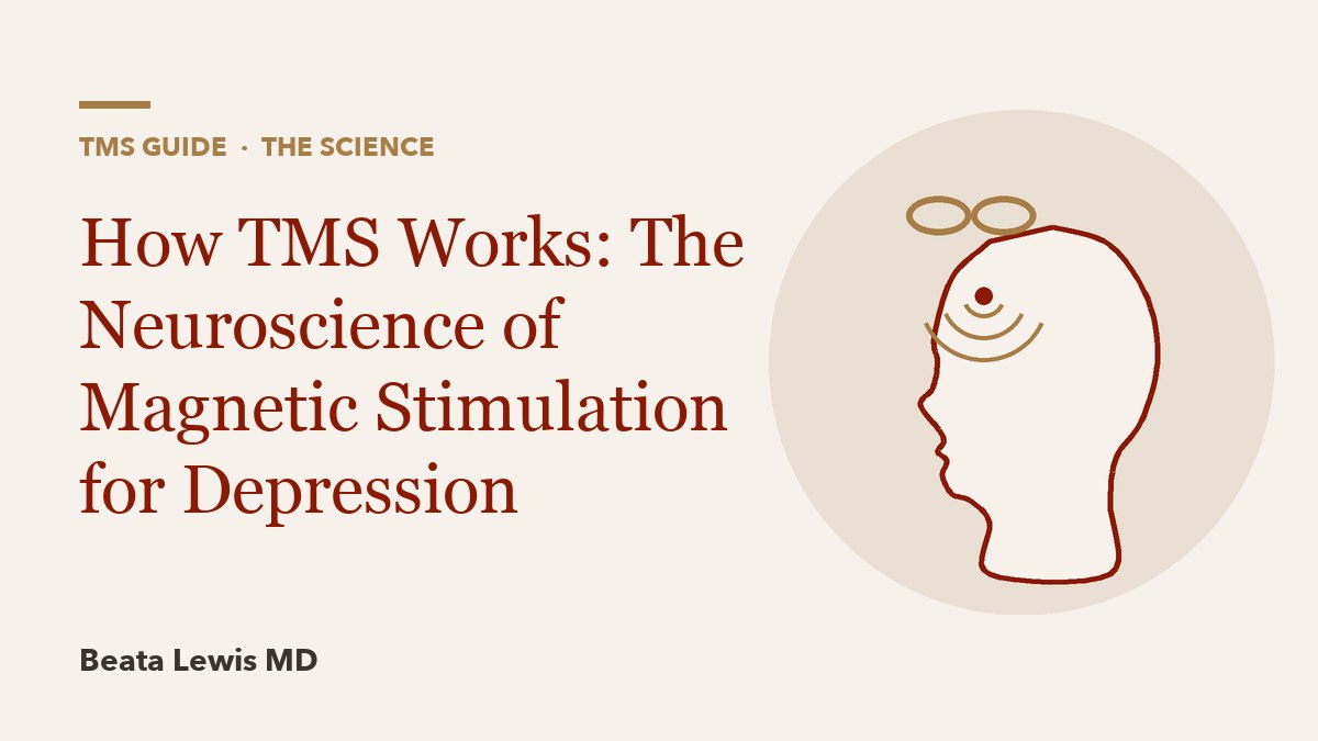
TMS works by turning a brief, strong magnetic pulse into a small electric current inside the brain. A coil held against the scalp produces a rapidly changing magnetic field that passes through the skull and induces enough current in the cortex beneath to make neurons fire. FDA's device regulation describes it in those terms: pulsed magnetic fields "of sufficient magnitude to induce neural action potentials in the prefrontal cortex" to treat major depression "without inducing seizure".
For depression, the pulses are aimed at the left dorsolateral prefrontal cortex (DLPFC). The antidepressant effect does not seem to come from that patch of cortex alone. Repeated stimulation over several weeks appears to change how the DLPFC communicates with deeper mood-regulating regions, especially the subgenual anterior cingulate cortex, through plasticity-like changes in synapses. Each link in that chain has supporting evidence, and each still has open questions, which this guide covers in order.
Step 1: From a magnetic pulse to an electric current
The physics is Faraday's law of electromagnetic induction. When a large current is switched through a copper coil for a fraction of a millisecond, it creates a magnetic field that rises and collapses very quickly. A changing magnetic field induces an electric field in any conductor nearby, and brain tissue conducts. The skull and scalp barely impede a magnetic field, which is why TMS can reach the cortex without electrodes, anesthesia or surgery.
Two features of the physics shape everything clinicians do with TMS:
- The induced field weakens quickly with depth. Standard coils mainly reach the outer cortex. In a 2005 comparison, a figure-8 coil could activate the motor cortex from about 2 cm away, while the deeper-reaching H-coil could do so from 5.5 cm.
- Depth costs focus. A simulation study of 50 coil designs found that every coil followed a depth-focality tradeoff: coils that reach deeper "cannot be as focal as more superficial coils." This is the core difference between standard and deep TMS with an H-coil.
The magnetic field itself is not the treatment. It is the delivery mechanism for an electric field, and the electric field is what acts on neurons.
Step 2: What a pulse does to a neuron
Neurons keep a voltage difference across their membranes. When the induced electric field is strong enough, it depolarizes axons in the stimulated area past threshold and they fire an action potential, the same all-or-none signal neurons use to communicate. Each pulse therefore produces a synchronized burst of real neural activity, which then travels along the connections of the neurons that fired.
How strong is strong enough differs from person to person, which is why every course starts by measuring the motor threshold. The coil is placed over the motor cortex, and the clinician finds the lowest intensity that moves the fingers, judged by visual inspection, sometimes with EMG. Treatment intensity is then set as a percentage of that number. In the trials that led to FDA clearance, the standard 10 Hz protocol ran at 120% of motor threshold. The reasoning is practical: the motor cortex gives a visible readout, while the prefrontal cortex does not. Distance matters too. The prefrontal target can sit further from the coil than the motor cortex, so some systems adjust for it: the SAINT system applies a depth correction for the distance between motor cortex and the frontal target, and its published protocol used 90% of resting motor threshold adjusted for cortical depth. The guide to motor threshold and dosing covers why the threshold is re-measured and what shifts it.
Step 3: Why the left dorsolateral prefrontal cortex
The early researchers chose the left DLPFC for a reason. An early pilot of daily left prefrontal TMS for depression, published in 1995 in six medication-resistant inpatients, cited "converging evidence" for "hypofunction of the left prefrontal cortex in depression". The DLPFC is part of the brain's executive control system, it sits close to the surface where a coil can reach it, and it is wired to deeper emotional circuits.
Finding the exact spot is harder than it sounds. The traditional "5 cm rule" places the coil 5 cm forward of the motor hand area. When researchers checked this method with neuronavigation in 22 people, it hit the intended DLPFC region in only 7; in the other 15 the coil sat further back or higher. A controlled 1996 study in The Lancet strengthened the case for this target: in a crossover design with sham and other cortical sites as controls, left DLPFC stimulation produced a significant drop in depression scores in 17 patients with medication-resistant psychotic depression. Newer methods include the Beam F3 skull measurement and MRI-guided neuronavigation, explained in the article on finding the TMS target.
Step 4: Network effects and the subgenual cingulate
The leading explanation for why prefrontal TMS treats depression centers on a deeper region called the subgenual anterior cingulate cortex (sgACC), which is involved in negative mood and is often overactive in depression.
In 2012, Fox and colleagues analyzed resting-state fMRI from 98 healthy people and replicated the finding in 13 depressed patients. DLPFC sites with better reported clinical results in past studies were those whose activity was more strongly "anticorrelated" with the sgACC; when one rose, the other tended to fall. The implication is that TMS may help by working through the DLPFC's connection to the sgACC, not just by exciting the DLPFC.
A 2014 imaging study before and after a 5-week TMS course in 17 depressed patients supported this. TMS "normalized depression-related subgenual hyperconnectivity" in the default mode network, a set of regions linked to rumination and self-focused thought, and baseline subgenual connectivity predicted who improved. It also induced anticorrelated connectivity between the DLPFC and medial prefrontal nodes of that network, while leaving connectivity within the executive network itself unchanged. In other words, the measurable change was in how the stimulated control region related to the rumination network, not simply more activity at the coil site.
The model is influential, but it has not yet produced consistent clinical gains. When connectivity-based targeting was tested in practice:
- The large BRIGhTMIND trial (2024) compared connectivity-guided iTBS with MRI-navigated standard 10 Hz rTMS in 255 patients and found no difference over 26 weeks.
- A 2026 trial of 40 adults found that connectivity-based targeting of accelerated TMS beat scalp-based targeting, with a median MADRS reduction of 24 vs 18 points.
- A 2026 trial of 123 patients found an early advantage for connectivity-guided targeting that was no longer significant by week 12.
The fair summary is that the sgACC network model is the best current explanation, and personalized targeting built on it is promising but not settled.
Step 5: Plasticity, and why frequency matters
A single pulse produces a brief burst of activity and nothing lasting. Repeated pulses at certain rhythms produce changes that outlast the stimulation, and those lasting changes are thought to drive the treatment effect. Most of what is known comes from motor-cortex experiments in healthy volunteers, where the effect can be measured as a change in the size of the muscle response to a test pulse.
| Pattern | What it did in motor-cortex studies | Clinical use in depression |
|---|---|---|
| High-frequency rTMS (5 Hz or higher) | Rapid-rate trains were followed by a period of increased excitability, attributed to "a transient increase in the efficacy of excitatory synapses" | 10 Hz to the left DLPFC, the original FDA-cleared protocol |
| Low-frequency rTMS (1 Hz or lower) | 0.9 Hz for 15 minutes reduced motor responses by an average of 19.5%, a decrease the authors compared to long-term depression (LTD) | Right DLPFC, rated a first-line protocol by CANMAT 2023 |
| Intermittent theta burst (iTBS) | Facilitatory; the original theta-burst study reported a "long-lasting, and powerful effect" on motor cortex physiology after 20 to 190 seconds of theta-burst stimulation | Left DLPFC in about 3 minutes per session |
| Continuous theta burst (cTBS) | Suppressive | Mainly research |
Two findings link these effects to synaptic plasticity. First, the low-frequency suppression resembles LTD, a weakening of synapses described in animal studies. Second, in a placebo-controlled study, memantine, a drug that blocks NMDA glutamate receptors, abolished both the facilitating effect of iTBS and the suppressive effect of cTBS. The authors concluded the after-effects "are likely to involve plasticity like changes at synaptic connections". NMDA receptors are central to long-term potentiation (LTP), the best-studied cellular basis of learning, which is why TMS effects are often called "LTP-like".
Two cautions apply. The prefrontal cortex is not the motor cortex, and responses vary widely between people, so "high frequency excites, low frequency inhibits" is a rule of thumb, not a law. And 2026 meta-analyses disagree on whether higher doses help: one found that more total pulses went with better outcomes in accelerated protocols, while another found "no significant linear dose-response relationships" across sham-controlled trials.
Why it takes many sessions
If TMS works by gradually reshaping circuits, a single session should not be enough, and the trial history agrees. The standard NeuroStar course is 5 days a week for 6 weeks plus a 3-week taper, 36 sessions in all. Consensus guidance tells patients that "a standard acute course of 20 to 30 treatment sessions over 6 weeks will very likely be needed" to match trial results. Even in the 1995 pilot, the two patients with clear benefit improved "progressively over the course of several weeks".
The open question is whether sessions need to be a day apart. Accelerated protocols deliver several sessions a day; across 59 studies in a 2026 meta-analysis the gap between sessions ranged from 10 to 480 minutes (median 50), and the analysis found intermediate spacing of about 30 to 75 minutes outperformed shorter or longer gaps. The accelerated TMS guide and the article on theta burst stimulation explain how those schedules are built and how strong the evidence is.
What TMS does not do
- It does not induce a seizure. That is the basic difference from ECT, which deliberately produces a controlled seizure under general anesthesia; see TMS vs ECT. An unintended seizure during TMS is a rare, labeled risk, discussed in TMS safety and seizure risk.
- It does not deliver a drug into the bloodstream. Its effects start locally under the coil and spread through connected networks, so its side-effect profile is mostly local: scalp discomfort and headache are the most common complaints.
- It does not work for everyone. Sham stimulation in trials produces sizable improvement on its own, a pooled effect size of about 0.8 across 61 trials, and active treatment has to beat that. The article on response vs remission explains how to read the numbers.
What the mechanism means for patients
The biology explains several practical features of a TMS course. Dose is individualized because motor thresholds differ. Coil placement matters because the target is a network hub, not a fixed scalp landmark. Consistent attendance matters because the effect builds through repeated stimulation. Sleep deprivation, alcohol withdrawal and certain metabolic problems are asked about before sessions because international safety guidelines list them as factors that lower seizure threshold. Researchers are still working out who will respond, a topic covered in predictors of TMS response.
The full set of guides is in the TMS guide. For an overview of TMS as a treatment option and how it fits into psychiatric care, see the TMS therapy page.
Frequently asked questions
Is the magnetic field itself what treats depression?
No. The magnetic field is the delivery system. It passes through the skull and induces a small electric current in the cortex, and that current makes neurons fire. The lasting antidepressant effect is thought to come from repeated firing reshaping how prefrontal and deeper mood circuits communicate.
How deep into the brain does TMS reach?
Standard figure-8 coils mainly reach the outer cortex directly. H-coils used for deep TMS reach further but stimulate a broader area, because every coil design trades depth for focus. Deeper structures such as the subgenual cingulate are thought to be influenced indirectly, through their connections with the stimulated cortex.
Does TMS change brain structure or just brain activity?
The best evidence is for changes in activity and connectivity, plus plasticity-like changes at synapses that depend on NMDA glutamate receptors. Claims that TMS "rewires" or "grows" the brain in a visible structural way go beyond what the clinical research shows.
Why is the left side of the brain stimulated?
Early researchers pointed to evidence of reduced activity in the left prefrontal cortex in depression, and high-frequency stimulation of the left DLPFC became the standard cleared protocol. Low-frequency stimulation of the right DLPFC is also used, and guidelines rate it as an evidence-based option.
Is TMS the same as ECT at a lower dose?
No. ECT passes current through the head to produce a controlled seizure under anesthesia. TMS uses magnetic induction to stimulate a targeted area without causing a seizure, and patients are awake and can drive afterward.
Sources
- 21 CFR 882.5805 Repetitive transcranial magnetic stimulation system. eCFR (U.S. Government Publishing Office), 2026. ecfr.gov
- K231926 510(k) Summary: NeuroStar Advanced Therapy System. U.S. FDA, 2024. accessdata.fda.gov
- K220177 510(k) Summary: Magnus Neuromodulation System with SAINT Technology. U.S. FDA, 2022. accessdata.fda.gov
- Daily left prefrontal transcranial magnetic stimulation therapy for major depressive disorder: a sham-controlled randomized trial. Archives of General Psychiatry, 2010. pubmed.ncbi.nlm.nih.gov
- Efficacy and safety of transcranial magnetic stimulation in the acute treatment of major depression: a multisite randomized controlled trial. Biological Psychiatry, 2007. pubmed.ncbi.nlm.nih.gov
- Transcranial magnetic stimulation in therapy studies: examination of the reliability of 'standard' coil positioning by neuronavigation. Biological Psychiatry, 2001. pubmed.ncbi.nlm.nih.gov
- Efficacy of transcranial magnetic stimulation targets for depression is related to intrinsic functional connectivity with the subgenual cingulate. Biological Psychiatry, 2012. pubmed.ncbi.nlm.nih.gov
- Connectivity-guided intermittent theta burst versus repetitive transcranial magnetic stimulation for treatment-resistant depression: a randomized controlled trial. Nature Medicine, 2024. pubmed.ncbi.nlm.nih.gov
- Connectivity- vs Scalp-Based Targeting of Accelerated Transcranial Magnetic Stimulation for Depression: A Randomized Clinical Trial. JAMA Psychiatry, 2026. jamanetwork.com
- Individualized Connectivity-Guided Versus Conventional Targeting of Accelerated Theta-Burst Stimulation in Depression: A Randomized, Double-Blind, Parallel-Design Trial. American Journal of Psychiatry, 2026. pubmed.ncbi.nlm.nih.gov
- Theta burst stimulation of the human motor cortex. Neuron, 2005. pubmed.ncbi.nlm.nih.gov
- K173620 510(k) Summary: MagVita TMS Therapy System w/Theta Burst Stimulation. U.S. FDA, 2018. accessdata.fda.gov
- Canadian Network for Mood and Anxiety Treatments (CANMAT) 2023 Update on Clinical Guidelines for Management of Major Depressive Disorder in Adults. Canadian Journal of Psychiatry, 2024. pmc.ncbi.nlm.nih.gov
- Dose-response associations of total pulse dose and intersession interval with clinical outcomes in accelerated rTMS for major depressive episodes: A systematic review and meta-analysis. Brain Stimulation, 2026. pubmed.ncbi.nlm.nih.gov
- Protocol-level moderators of antidepressant efficacy in prefrontal rTMS: A sham-controlled meta-analysis of randomized trials. Journal of Affective Disorders, 2026. pubmed.ncbi.nlm.nih.gov
- Consensus Recommendations for the Clinical Application of Repetitive Transcranial Magnetic Stimulation (rTMS) in the Treatment of Depression. Journal of Clinical Psychiatry, 2018. pmc.ncbi.nlm.nih.gov
- A systematic review and meta-analysis on placebo response to repetitive transcranial magnetic stimulation for depression trials. Progress in Neuro-Psychopharmacology and Biological Psychiatry, 2018. pubmed.ncbi.nlm.nih.gov
- Transcranial magnetic stimulation in the treatment of major depressive disorder: a comprehensive summary of safety experience from acute exposure, extended exposure, and during reintroduction treatment. Journal of Clinical Psychiatry, 2008. europepmc.org
- Safety and recommendations for TMS use in healthy subjects and patient populations, with updates on training, ethical and regulatory issues: Expert Guidelines. Clinical Neurophysiology, 2021. europepmc.org
- Stanford Accelerated Intelligent Neuromodulation Therapy for Treatment-Resistant Depression. American Journal of Psychiatry, 2020. pubmed.ncbi.nlm.nih.gov
- Rapid-rate transcranial magnetic stimulation of left dorsolateral prefrontal cortex in drug-resistant depression. The Lancet, 1996. pubmed.ncbi.nlm.nih.gov
- Transcranial magnetic stimulation of deep brain regions: evidence for efficacy of the H-coil. Clinical Neurophysiology, 2005. pubmed.ncbi.nlm.nih.gov
- Electric field depth-focality tradeoff in transcranial magnetic stimulation: simulation comparison of 50 coil designs. Brain Stimulation, 2013. pubmed.ncbi.nlm.nih.gov
- Daily repetitive transcranial magnetic stimulation (rTMS) improves mood in depression. NeuroReport, 1995. pubmed.ncbi.nlm.nih.gov
- Default mode network mechanisms of transcranial magnetic stimulation in depression. Biological Psychiatry, 2014. pubmed.ncbi.nlm.nih.gov
- Responses to rapid-rate transcranial magnetic stimulation of the human motor cortex. Brain, 1994. pubmed.ncbi.nlm.nih.gov
- Depression of motor cortex excitability by low-frequency transcranial magnetic stimulation. Neurology, 1997. pubmed.ncbi.nlm.nih.gov
- The after-effect of human theta burst stimulation is NMDA receptor dependent. Clinical Neurophysiology, 2007. pubmed.ncbi.nlm.nih.gov
Educational information, not medical advice. Whether TMS or any treatment fits you is a decision to make with a psychiatrist who knows your history.
The information provided on this blog is for educational and informational purposes only. It is not intended to be a substitute for professional medical advice, diagnosis, or treatment. Always seek the advice of your physician or other qualified health provider with any questions you may have regarding a medical condition.

A Message From Dr. Lewis If you’ve been struggling with depression or anxiety, I want you to know something important:

The Conversation That Surprised Me A few years ago, I had a patient named Greg who’d been on escitalopram (Lexapro)


You’re Not Meant to Go Through Life Alone In our increasingly connected yet often isolating world, you might sometimes feel